
How Sedation Monitoring Works at the Dentist
Learn how dental teams continuously track vital signs during sedation, the equipment used, and what patients can expect for a safe visit.
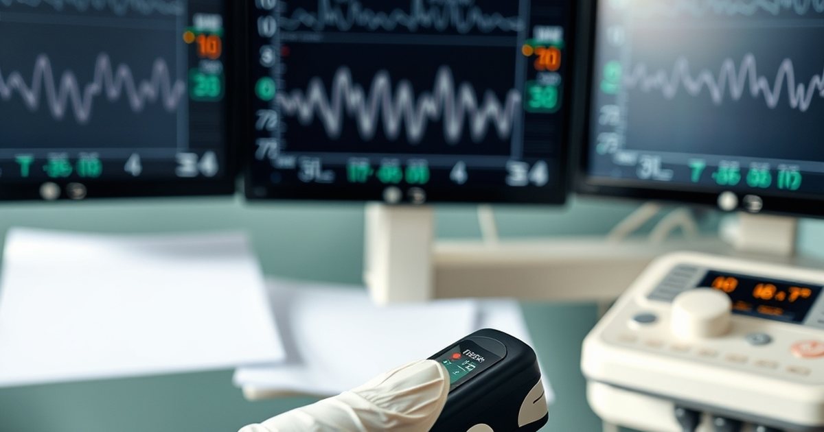
During dental sedation, a trained clinician continuously records heart rate, blood pressure, oxygen saturation, and breathing using pulse oximetry, capnography, and automated cuffs to detect.
At a Glance
- Primary goal: Detect physiologic changes early
- Core devices: Pulse oximeter, capnograph, automated BP cuff
- Monitoring frequency: Continuous, real‑time display
- Team role: Dedicated monitor or anesthesia‑trained provider
At a Glance
- Primary goal
- Detect physiologic changes early
- Core devices
- Pulse oximeter, capnograph, automated BP cuff
- Monitoring frequency
- Continuous, real‑time display
- Team role
- Dedicated monitor or anesthesia‑trained provider
Pulse Oximetry vs Capnography
| Feature | Pulse Oximetry | Capnography |
|---|---|---|
| What it measures | Arterial oxygen saturation (SpO₂) and pulse rate | End‑tidal carbon dioxide (ETCO₂) and respiratory rate |
| Typical clinical use | Standard for all sedation levels | Added for moderate or deep sedation |
| Invasiveness | Non‑invasive finger or earlobe sensor | Non‑invasive nasal cannula or airway adapter |
| Data frequency | Continuous waveform every few seconds | Continuous numeric and waveform display |
| Alarm thresholds | SpO₂ below 90 % triggers alarm | ETCO₂ above 50 mmHg or apnea >10 s triggers alarm |
During dental sedation, a trained clinician continuously records heart rate, blood pressure, oxygen saturation, and breathing using pulse oximetry, capnography, and automated cuffs to detect changes instantly. This real‑time oversight is the cornerstone of safe sedation practice in the dental office.
What Is Sedation Monitoring?
Sedation monitoring is the systematic, continuous observation of a patient’s physiologic parameters while sedative medications are active. The mechanism relies on non‑invasive sensors that feed data to a bedside display, allowing the monitoring provider to recognize trends or abrupt shifts before they become clinically significant. A pulse‑oximeter shines light through a fingertip or earlobe to calculate arterial oxygen saturation and pulse rate, while a capnograph samples exhaled gas through a nasal cannula to measure end‑tidal carbon dioxide and respiratory rate. An automated cuff inflates at set intervals to capture systolic and diastolic pressures, and ECG leads may be added for deeper levels of sedation. For the patient, the experience is largely passive: a gentle pressure on the finger, a soft nasal prong, and the occasional squeeze of the cuff, all while they remain relaxed and often unaware of the continuous data stream that safeguards their wellbeing.
Monitoring Methods Compared
| Aspect | Pulse Oximetry | Capnography |
|---|---|---|
| What it measures | Arterial oxygen saturation (SpO₂) and pulse rate | End‑tidal carbon dioxide (ETCO₂) and respiratory rate |
| Typical clinical use | Standard for all sedation levels | Added for moderate or deep sedation |
| Invasiveness | Non‑invasive finger or earlobe sensor | Non‑invasive nasal cannula or airway adapter |
| Data frequency | Continuous waveform every few seconds | Continuous numeric and waveform display |
| Alarm thresholds | SpO₂ below 90 % triggers alarm | ETCO₂ above 50 mmHg or apnea >10 s triggers alarm |
How Sedation Monitoring Is Performed
- Pre‑procedure baseline – The monitoring clinician records resting heart rate, blood pressure, SpO₂, and ETCO₂ before any sedative is given. The patient feels a brief, painless cuff inflation and a cool sensor on the fingertip while sitting quietly.
- Sensor placement – A pulse‑oximeter probe is attached to a finger or earlobe; a nasal cannula or airway adapter connects to the capnograph; an automated cuff is positioned on the upper arm; ECG leads may be added for deeper sedation. The patient notices a light adhesive strip on the finger and a soft tube resting near the nostrils, both barely noticeable.
- Continuous display – All waveforms and numeric values appear on a single monitor screen visible to the dedicated provider throughout the case. The patient sees only the dental team working; the monitor remains out of sight, silently tracking each breath and heartbeat.
- Scheduled documentation – Values are entered into the clinical record at least every five minutes, with additional entries during induction, dose changes, or any alarm. The patient experiences no interruption; the clinician simply notes numbers while the procedure continues.
- Alarm response – If a parameter crosses a preset threshold, the clinician follows a stepwise protocol: verify the reading, adjust oxygen or ventilation, modify sedation depth, and prepare for advanced airway support if needed. The patient may feel a brief increase in oxygen flow or a gentle verbal cue to take a deeper breath, but the response is swift and usually unnoticed.
- Post‑procedure observation – Monitoring continues until the patient meets discharge criteria — stable vitals, adequate oxygenation, and return to baseline mental status. The patient rests in a recovery chair while the same sensors remain in place, feeling a gradual return to full alertness.
Key Numbers from Guidelines
- The ADA sedation guidelines recommend maintaining SpO₂ at or above 90 % throughout the procedure.
- Capnography alarms are typically set to trigger when ETCO₂ exceeds 50 mmHg or when no breath is detected for more than 10 seconds.
- Blood‑pressure readings are documented at minimum every five minutes, with more frequent checks during medication titration.
- A dedicated monitoring provider is required for any sedation level beyond minimal anxiolysis.
- Post‑sedation observation continues until the patient’s vital signs have remained stable for at least 15 minutes.
Patient Selection, Risks, and Aftercare
Who Is a Good Candidate?
Patients who are in generally good health, have a clear airway, and can cooperate with simple instructions tend to tolerate dental sedation well. Those with controlled chronic conditions such as hypertension or diabetes, provided they are stable and medication‑compliant, are also appropriate candidates. The dental team evaluates medical history, current medications, and the planned procedure length to match the sedation depth to the individual’s needs.
Contraindications and Airway Considerations
Certain situations raise the risk of complications and may lead the provider to defer or modify sedation. An unsecured or anatomically difficult airway, severe obstructive sleep apnoea, or a history of malignant hyperthermia are relative contraindications. Pregnancy, especially in the first and third trimesters, warrants careful risk‑benefit discussion because sedative agents can cross the placenta. Active respiratory infection, uncontrolled asthma, or recent upper‑airway surgery also increase the likelihood of airway compromise during sedation.
Risks and Team Management
Even with continuous monitoring, sedation carries inherent risks such as transient hypoxemia, hypotension, or brief periods of apnea. The monitoring clinician mitigates these by maintaining supplemental oxygen, adjusting drug infusion rates, and using verbal or tactile stimulation to encourage spontaneous breathing. Emergency equipment — bag‑valve‑mask, suction, and reversal agents — is kept immediately accessible, and the team rehearses a standardized rescue protocol before each case. Early detection through capnography and pulse oximetry allows intervention long before a serious event develops.
First 24 Hours After Sedation
After discharge, the patient should remain under the supervision of a responsible adult for the remainder of the day. Drowsiness, mild nausea, and a dry mouth are common and usually resolve within a few hours. The patient is advised to avoid driving, operating machinery, making legal decisions, or consuming alcohol for at least 24 hours. Clear fluids can be resumed once the gag reflex has returned, progressing to light meals as tolerated. Any persistent vomiting, difficulty breathing, unusual confusion, or fever should prompt an immediate call to the dental office or emergency services.
When to See a Dentist
- Persistent nausea or vomiting after the appointment
- Difficulty breathing or shortness of breath that does not resolve quickly
- Unusual drowsiness that prevents normal activity several hours later
- Signs of allergic reaction such as rash, swelling, or itching
- Uncontrolled bleeding from the treatment site
- Fever above 101 °F (38.3 °C) within 24 hours
- Severe pain not relieved by prescribed medication
FAQ
What equipment is used for sedation monitoring?
A pulse oximeter, capnograph, automated blood‑pressure cuff, and often an ECG lead set are standard. Each device provides a real‑time stream of data that the monitoring clinician watches throughout the procedure.
Who is responsible for monitoring during sedation?
A qualified provider — often the dentist with sedation training, a dental anesthesiologist, or a certified registered nurse anesthetist — is assigned solely to observe the monitors and respond to any changes.
How often are vital signs recorded?
Values are displayed continuously and documented at minimum every five minutes, with more frequent charting during induction, emergence, or any event that deviates from baseline.
What happens if a reading falls outside normal range?
The monitoring clinician follows a predefined protocol: adjust oxygen flow, modify sedation depth, stimulate breathing, or, if needed, activate emergency resuscitation measures.
Can patients request a specific monitoring method?
Patients may discuss preferences with their provider, but the final monitoring plan follows evidence‑based guidelines that prioritize safety for the chosen sedation level.
Is sedation monitoring required for all levels of sedation?
Guidelines mandate continuous monitoring for minimal, moderate, and deep sedation. The specific devices required increase with the depth of sedation.
Sources
- https://www.ada.org/-/media/project/ada-organization/ada/ada-org/files/resources/practice/guidelines/sedation_anesthesia_guidelines.pdf
- https://www.asahq.org/madeforthismoment/anesthesia-101/types-of-anesthesia/
- https://www.ncbi.nlm.nih.gov/books/NBK470310/
This article is for general information and is not medical advice.
Frequently Asked Questions
What equipment is used for sedation monitoring?
Who is responsible for monitoring during sedation?
How often are vital signs recorded?
What happens if a reading falls outside normal range?
Can patients request a specific monitoring method?
Is sedation monitoring required for all levels of sedation?
More from the blog
Medical Disclaimer: The clinical information provided on Sedation Dentistry is intended strictly for educational and informational purposes. It does not replace an individual assessment, a consultation, or advice from a licensed dentist, anaesthesiologist or physician. Sedation decisions depend on your airway, medications and medical history. In an emergency, call 911.